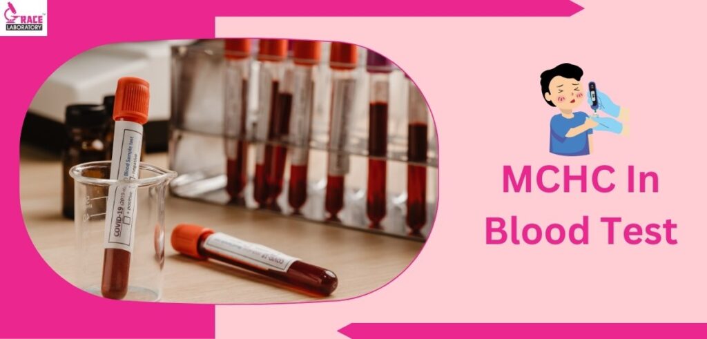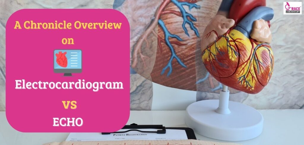
“ A Healthy Heart Shines in Bonniest- Beauty”
Electrocardiograms and Echocardiograms are the most common noninvasive cardiology tests that monitor your heart activity and heart-related disease. These are standard and graphic electrical impulses that form an electrical pattern usually in amplitude wave-form to check for any heart- abnormalities. Although these two common diagnostic tools check the heart and identify valuable information regarding your electrical heart activities and its functioning, they have different methodologies and purposes.
In conventional apprehensions; we understand that both these techniques are important in recording the sequence of heart graphical stimulus; however, this article will give you a comprehensive and complementary understanding of Electrocardiogram vs ECHO. As the name suggests; an Electrocardiogram stands for ECG; the process of recording the graph voltage of the heart’s electrical impulse using electrodes whereas an ECHO is an ultrasound that stands for sound to detect fatal cardiac abnormalities. So if you are consistently experiencing chest- pain, woozy and dizziness, heart pounding, or throbbing ( Heavy Palpitations); Grace Laboratory Doctors will suggest an ECHO Test; after the results, if there are any warning signs then we perform an Electrocardiogram Test.
The Difference between an Echocardiogram and Electrocardiogram
However, regardless of age and gender, cardiovascular problems and strokes are leading problems among us; therefore The Difference between an Echocardiogram and Electrocardiogram has become an important consideration in understanding it. Now that we know what Electrocardiogram and ECHO are; below is a distinctive summary of- Electrocardiogram vs ECHO.
- Echocardiogram- ECHO primarily provides complete and detailed information about structural heart- disease and congenital abnormalities through an ultrasound. It produces a high-quality sound wave about the chambers of your heart by producing a real-time multi-dimensional image of the heart valves. The high-emitted sound- waves are generated by placing the electronic device: transducer on to the chest. During the ultrasound; ECHO records the electrical signals on the monitor and the doctor can see the live footage of the muscular- heart. This test gives a mechanical rhythm of the heart waves.
- Electrocardiogram- ECG or EKG outlines the electrical heart impulses as they beat in a waveform using electrodes. An Electrocardiogram test of the valve and endocarditis congenital heart diseases is performed during Artery Disease, Heart Failures, and heart disease treatments such as pacemakers. These are more or less painless tests depending on the heart’s autonomy and heart condition.
What Is An Electrocardiogram And How Does It Work?
As we know Electrocardiogram( EKG or ECG) is a signal-averaged machine test monitored over a portable ambulatory electrocardiography device that records the irregularities of heartbeat changes for 24 hours. This note will explain to you What Is An Electrocardiogram And How Does It Work?
The circulatory heart and cardiovascular system are vital organs in our human body. Moreover, the prevalence of cardio-coronary diseases has become a resolute condition. An Electrocardiogram test will help the doctor to record Coronary artery disease( CAD) and complications through electrical signals.
How Does It Work?– During the test; the cardiologist will take at least 10 minutes to place the 12 small- sticky electrodes or sensors on the surface of your chest wall skin, legs, and arms to diagnose the Hypertrophic cardiomyopathy( HCM) and other heart complications. These electrodes detect electrical heart activity and impulses (arrhythmias) of the patient over time and transmit the data to an implantable cardiac device.
Generally, there are many types of ECG tests depending on the heart condition and it is always recommended to come empty stomach before an EKG or ECG machine test. Furthermore; you can collect the EKG test results within seven days.
What’s an Echocardiogram And How Does It Work?
Echocardiogram is slightly different from Electrocardiogram tests that evaluate the rhyme of the heart. Echocardiogram is the process of mechanical Carotid ultrasound and Electrocardiogram measures the electrical heart- impulse. Our heart is an indispensable organ that includes heart walls, heart chambers, heart valves, and blood vessels. This medical test is required when you have chest- pain or any other congenital heart aterary defects.
How Does It Work?- ECHO tests are mainly of two types- TTE(Transesophageal Electrocardiogram) and TEE ( Transesophageal Echocardiogram). There are certain steps taken into consideration before performing the Echocardiograms. The sonographer or the cardiologist will initially apply the gel in the front of the chest wall and use an electrical device: a transducer to produce an ultrasound wave image to investigate the chest pain and shortness of breathe. The Holter monitor examines the track of the heartbeats and identifies the potential cause of ruptured blood vessels that cause heart attack stroke.
So Echocardiograms are a common diagnostic procedure of high-frequency ultrasound when you experience uncomfortable chest pain.
How Are The Tests Done?
Echocardiogram and Electrocardiogram diagnostic tests are important tests that account for the heart rhythm and recognize the cardiovascular conditions of the heart. The purpose of these medical tests showcase the heart rhythm through the conjunction of mechanical and electrical systems. Moreover, this note will elucidate how these tests manage the cardiac condition and How Are The Tests Done.
As we know both heart tests are common, simple, safe, and non-invasive specialized tests to study the abnormalities in the structure of the heart. These tests are performed in hospitals by a practicing cardiologist and physician. During Transthoracic ECHO and EKG or ECG procedures; you are required not to eat before 8 hours.
- A standard ECHO is conducted by applying lubricating- jelly-like gel on an ultrasound- wand. The doctor or the sonographer testifies the damaged heart tissue, the thickness of the heart valves and chambers along any blood clots through the produced image on the transducer.
- As an Echocardiogram cannot check and record the electrical problems; therefore EGC diagnostic test is supervised where sensors like 12 lead electrodes are placed over the chest-skin to measure the rhythmic waves and impulses of the heart. You are now asked to breathe in where the Holter monitor records the information and transfers(analog to digital) the electricity activity on the paper or monitor.
After the test; if your heart specialist witnesses some complications and problems then you may be referred to more tests depending on the heart situation.
When Do You Need Them?
The phenomenon of recording echo and electrical heart rhyme becomes the first choice when it comes to the heart. Moreover; the ECHO and ECG cardio management record system remains an integral and elucidative tool for heartbeat readings.
- The Purpose of an Echocardiogram- In context to the heart’s structural abnormalities and disease- CHD( Congenital heart disease), heart failure, blocking of the heart’s arteries due to cholesterol deposits, capturing of the heart muscles and irregular blood flow pattern from left to right side heart atrium or vice- versa resulting in stunts; ECHO is needed. Generally, this ultrasound medical procedure requires a minimum preparation to visualize the four-chambered heart anatomy.
- The Purpose of an Electrocardiogram- You need an ECG when you undergo severe chest pain, heart palpitation, uneven heartbeats, and Dyspnea- difficulty in breathing( suffocation). ECG medical tests are usually required to measure the tracing intervals of the TP, PR, and QRS segments of heart valves. The 12 ECG leads to understanding and determining the disturbance/ irregularities from P to S conduction electrical patterns of every heartbeat.
Electrocardiogram vs ECHO
Both these cardiovascular tools sound a bit similar; however, they serve different purposes. Now that we have deeply understood the functions of echo and electrocardiograms; find out The Difference between an Echocardiogram and Electrocardiogram in the tabulated form below.
Electrocardiogram | Echocardiogram |
It happens in some seconds only | The procedure might take an hour |
It uses electrodes to determine the asymmetry in the heart’s structure | It uses an echo/sound to provide detailed information on chambers, valves, and the blood flow. |
ECG can determine the heart’s blockage. | The test cannot record the heart blockage |
The medical test is responsible for bestowing details on cardiac rhythm | Echo examines the changes in the cardiac structure |
Towards the end, I would say that our heart is a central body engine that constantly maintains blood pressure and supplies oxygenated blood and nutrients. Hence, this fist-sized cardiovascular organ plays an important role in your life. So, in broad terms, if you are witnessing any heart-related conditions and consistent irregular behavior in your heart- waves you should immediately get examined for heart health.
Grace Laboratory based out in Vadodara, Gujarat is a leading multi-specialist blood test laboratory clinic for health- care 24*7 seamlessly with ultra-pleasant satisfaction and accurate patient results. Moreover, the renowned clinic has created so many successful accreditations with a mission-
- G – Growth for all stakeholders
- R – Rapid service with a human touch
- A – Accurate
- C – Customer satisfaction
- E – Ethical & Innovative
Go and request your schedule from https://gracelaboratory.com/ soon.